This article was published on: 11/11/22 4:48 AM
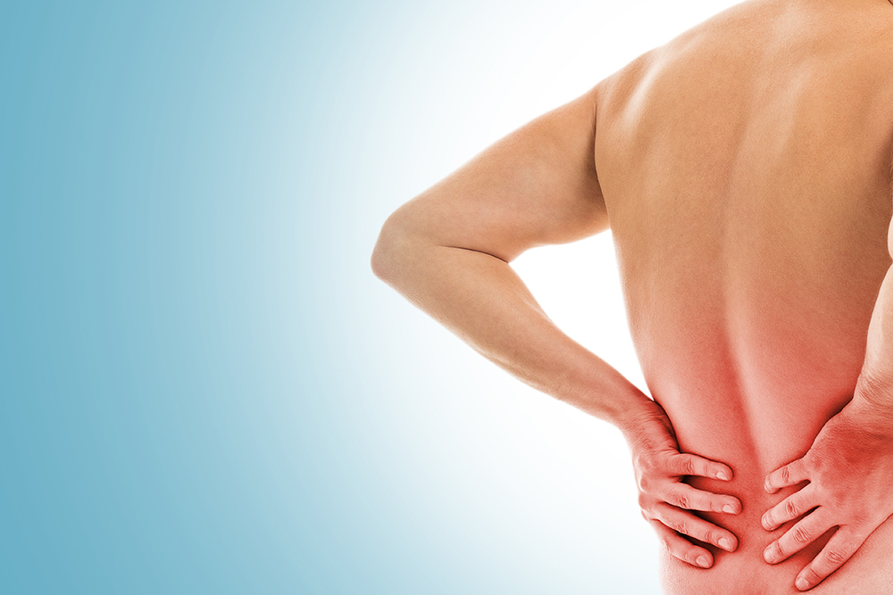
What is Anterior Knee Pain?
Anterior knee pain is the best way to define the painful condition that arises in or around the front and central aspect of the knee this pain occurs by injury or overuse. According to doctors, it is common for people who are in sports, adults, and women. It will get worse by worse when you will feel pain in your daily activities when you walk downstairs, squat while driving the car, and your knees are in a flexed position for a long time. Anterior knee pain also has some alternative names such as
What are the Causes of Anterior Knee Pain?
Anterior Knee Pain usually occurs when your knee cap does not move properly instead it rubs against the lower part of the thigh bone. This happens due to the abnormal position of the knee cap. Anterior Knee Pain can also be caused due to various problems such as the runner’s knee, lateral compression syndrome, chondromalacia of the patella, etc.
Some of the other causes of Anterior Knee Pain include structural abnormalities or damage, muscle weakness, overuse of the knee, patellar fracture, patellofemoral instability, etc.
According to doctors anterior knee pain is commonly found in people who are overweight, who had a dislocation, fracture or it can be any other injury related to the kneecap.
Some of the common possibilities of anterior knee pain are arthritis and synovial impingement/ plica syndrome
What are the Symptoms of Anterior Knee Pain?
When your ankle is moving towards or moving closer to the backside of your thigh you will feel some kind of grating or it can be grinding. Some sounds will occur like crackling/popping from your knees when you bend your knees or climb stairs.
You will feel pain behind or around the knee cap while performing activities such as wearing heels, climbing or going down the stairs, squatting, sitting for a long period in one position, etc.
What is the Differential Diagnosis of Anterior Knee Pain?
The anterior knee can be structurally organized into four layers:
Layer 1: Soft tissue
Soft tissue consists of skin, fascia, bursae, and subcutaneous fat. According to the doctors, bursitis is a usual pathology that can affect this layer.
What is Bursitis?
Bursae are synovial-lined sacs that make possible movement between soft tissues. Bursitis causes Swelling or thickening of the bursal wall resulting in inflammation of the bursa. The common causes of Bursitis include acute trauma, infection, inflammatory arthritis, or chronic microtrauma. There are 4 types of bursae in the anterior knee: the prepatellar, superficial infrapatellar, deep infrapatellar, and pes anserine bursae. The prepatellar bursa is the most common superficial Bursitis after olecranon bursitis is easily affected. Infrapatellar bursitis must be told apart from fat pad impingement syndrome.
Pes anserine bursitis can be known as a tribal stress fracture, chondral fracture, meniscal tear, or osteonecrosis.
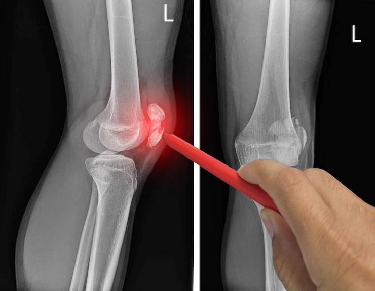
What is the Diagnosis of Bursitis?
Diagnosis of Bursitis depends upon your medical history along with the physical exam done by your healthcare provider. Radiography plays a very important role in finding out the history of the trauma along with any fracture or other bony abnormalities whereas if the bursal swelling is obscured by swelling due to joint effusionIf, ultrasound can be helpful. Hyperemia along with inflammation can be found through color doppler. Your doctor might even ask you to go for various blood tests or analysis of fluid from the bursa to identify the cause of the pain and swelling. Other tests that your doctor might order are MRI and X-ray.
Layer 2: Extensor Mechanism:
The extensor mechanism consists of the tibial tuberosity, quadriceps tendon, patella tendon, and patella. Excessive stress on these structures might result into jumper knee
What is the diagnosis of Extensor Mechanism?
Diagnosis of Extensor Mechanism injuries depends upon your medical history along with the physical exam done by your healthcare provider. There is a risk that the symptoms of injuries may come back in the future even after the diagnosis. Clinical evaluation, MRI, and X-rays can be taken to identify the cause of the problem. A physical examination can also be helpful in determining the structure that is causing pain. Your doctor might even ask you to come for a routine knee check up to know the progress.
Layer 3: Intracapsular or Extrasynovial:
The intracapsular layer is situated outside the joint capsule. This layer consists of a fat pad. Injury to the fat pad generally occurs due to the overuse of the intracapsular layer which results in bleeding and welling. There are three types of fat pad i.e The intracapsular infrapatellar fat pad, suprapatellar fat pad, and prepatellar fat pad.
What is the diagnosis of Intracapsular or Extrasynovial?
The diagnosis of fat pad injury is generally through a non-surgical process. Different types of methods that can be used to treat fat pad injury are rest, cold therapy, electrotherapy, patella taping, injection, and exercises. In rare cases, where the fat pad injury is not cured through the above methods, surgery is done.
Layer 4: Intraarticular
Intraarticular is situated in the interior of the joints. This layer consists of synovium and plica. Intraarticular.
What is the diagnosis of Intraarticular?
Plica syndrome is the only way to diagnose clinically. The predisposing causes, it can be found by imaging and other etiologies. This helps in assisting the doctor’s surgical planning. Radiographs and weight-bearing anteroposterior are also used to identify the cause of the problem.